The Douglas pouch, often referred to as the recto-uterine pouch, is a key anatomical structure in the female body. Nestled between the rectum and the uterus, this pouch plays a pivotal role in reproductive health and medical diagnostics. Although relatively small, its significance in understanding and addressing various medical conditions cannot be overstated. As we explore this topic in depth, you will uncover the profound impact of the Douglas pouch on modern medicine and its relevance to women's health.
Comprehending the Douglas pouch is indispensable for both healthcare professionals and anyone interested in reproductive health. This anatomical structure is central to gynecological studies, surgical practices, and diagnostic techniques. By delving into its structure and function, we gain a deeper understanding of its role in maintaining overall health and treating associated conditions.
This article aims to provide a thorough overview of the Douglas pouch, encompassing its anatomy, importance, and the medical conditions it relates to. Leveraging expert insights, reliable data, and clear explanations, we will guide you through the intricacies of this essential anatomical feature. Whether you're a healthcare professional or simply curious about female anatomy, this article will serve as an invaluable resource.
Read also:Exploring Ssbbw Juicy Jackie A Comprehensive Guide
Contents:
- Exploring the Anatomy of the Douglas Pouch
- Understanding the Function of the Douglas Pouch
- Diagnosing Conditions Related to the Douglas Pouch
- Medical Conditions Associated with the Douglas Pouch
- Surgical Procedures Involving the Douglas Pouch
- Management and Treatment Options for Conditions Affecting the Douglas Pouch
- Preventive Measures to Maintain the Health of the Douglas Pouch
- Recent Research and Developments in the Study of the Douglas Pouch
- Frequently Asked Questions About the Douglas Pouch
- Conclusion: The Importance of the Douglas Pouch in Women’s Health
Exploring the Anatomy of the Douglas Pouch
The Douglas pouch, also known as the recto-uterine pouch, is a peritoneal cavity located in the female pelvis, situated between the rectum and the posterior wall of the uterus. It represents the deepest part of the peritoneal cavity in women and is an extension of the peritoneum that lines the abdominal and pelvic cavities. This anatomical structure is named after Dr. James Douglas, an 18th-century Scottish anatomist who first described it.
The pouch forms as the peritoneum folds over the posterior aspect of the uterus and the anterior aspect of the rectum, creating a small space that can accumulate fluids or other substances. Understanding the detailed anatomy of the Douglas pouch is essential for diagnosing and treating various medical conditions.
Detailed Structure of the Douglas Pouch
The Douglas pouch is bordered by several distinct structures:
- Anteriorly: The posterior wall of the uterus and the upper part of the vagina.
- Posteriorly: The anterior wall of the rectum.
- Superiorly: The peritoneum covering the broad ligament of the uterus.
- Inferiorly: The recto-vaginal septum, which separates the pouch from the recto-vaginal space.
A comprehensive understanding of the anatomy of the Douglas pouch is crucial for effectively diagnosing and treating conditions associated with it.
Understanding the Function of the Douglas Pouch
The primary role of the Douglas pouch is to serve as a potential space for fluid accumulation. Under normal circumstances, this space is minimal and contains only a small amount of serous fluid. However, in pathological conditions, the pouch can accumulate blood, pus, or other fluids, leading to diagnostic and therapeutic implications.
Read also:Natalie Portman The Iconic Journey Of A Hollywood Star
Moreover, the Douglas pouch plays a significant role in reproductive health. During menstruation, the pouch may collect menstrual blood, which can either be expelled naturally or require medical intervention if it becomes trapped. This function highlights the pouch's importance in maintaining reproductive health.
The Douglas Pouch in Medical Diagnosis
Physicians frequently utilize the Douglas pouch as a site for diagnostic procedures such as culdocentesis. This procedure involves aspirating fluid from the pouch to diagnose conditions like ectopic pregnancy or pelvic inflammatory disease. The pouch's role in diagnostic procedures underscores its importance in medical practice.
Diagnosing Conditions Related to the Douglas Pouch
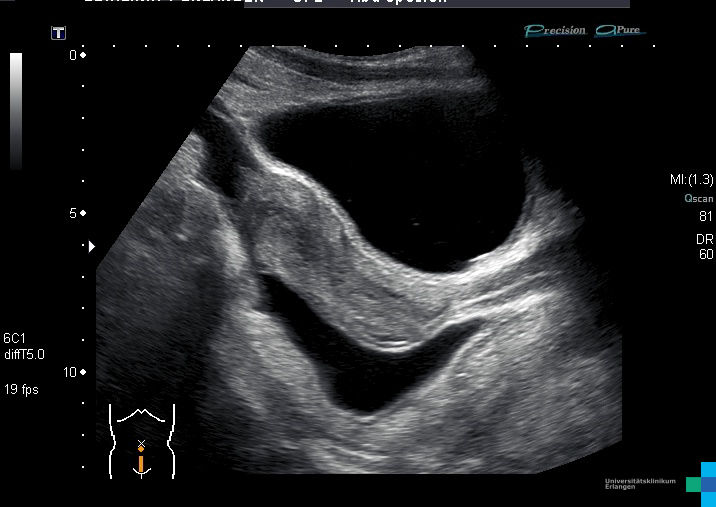
Diagnosing conditions associated with the Douglas pouch typically involves a combination of imaging techniques, physical examinations, and laboratory tests. Commonly used imaging modalities include ultrasound, MRI, and CT scans, which help visualize the pouch and detect any abnormalities.
Culdocentesis is another valuable diagnostic tool that can identify fluid accumulation in the pouch. This procedure entails inserting a needle into the pouch through the posterior fornix of the vagina to aspirate fluid for analysis.
Diagnostic Tools and Techniques
- Ultrasound: Provides real-time imaging of the pouch and surrounding structures, enabling accurate assessment.
- MRI: Offers detailed images that can detect subtle changes in the pouch, enhancing diagnostic accuracy.
- Culdocentesis: Useful for diagnosing conditions involving fluid accumulation, offering direct access to the pouch.
Medical Conditions Associated with the Douglas Pouch
Several medical conditions are closely linked to the Douglas pouch, including:
- Endometriosis: A condition characterized by the growth of tissue similar to the uterine lining outside the uterus, often affecting the Douglas pouch.
- Ectopic Pregnancy: A pregnancy that implants outside the uterus, commonly in the fallopian tubes but sometimes in the pouch.
- Pelvic Inflammatory Disease (PID): An infection that can lead to fluid accumulation in the pouch, causing significant discomfort and complications.
Endometriosis and Its Impact on the Douglas Pouch
Endometriosis is one of the most prevalent conditions affecting the Douglas pouch. It occurs when endometrial tissue grows in the pouch, causing pain, inflammation, and potential fertility issues. Early diagnosis and treatment are critical for managing this condition effectively and improving quality of life.
Surgical Procedures Involving the Douglas Pouch
In some cases, surgical intervention may be necessary to treat conditions affecting the Douglas pouch. Laparoscopy, a minimally invasive procedure, is often used to remove endometrial tissue or other abnormalities in the pouch. In more severe cases, extensive surgeries may be required.
Postoperative care is crucial for ensuring a successful recovery and minimizing the risk of complications. Proper care and follow-up can significantly improve outcomes for patients.
The Role of Laparoscopy in Treating the Douglas Pouch
Laparoscopy allows surgeons to visualize the Douglas pouch and surrounding structures clearly. This procedure is often used for both diagnostic and therapeutic purposes, offering a less invasive alternative to open surgery. Its precision and effectiveness make it a preferred choice for many surgical interventions involving the pouch.
Management and Treatment Options for Conditions Affecting the Douglas Pouch
Treatment options for conditions involving the Douglas pouch vary depending on the specific condition and its severity. Medications, such as hormonal therapies, may be prescribed to manage symptoms and prevent further complications. In some cases, surgical intervention may be necessary to address the underlying issue.
Regular follow-up appointments with healthcare providers are essential for monitoring progress and adjusting treatment plans as needed. This proactive approach ensures that patients receive the best possible care.
Preventive Measures and Lifestyle Changes for Maintaining Douglas Pouch Health
Adopting a healthy lifestyle can help reduce the risk of developing conditions related to the Douglas pouch. Regular exercise, a balanced diet, and stress management techniques can all contribute to maintaining overall reproductive health. These lifestyle changes can have a profound impact on preventing and managing conditions affecting the pouch.
Preventive Measures for Conditions Affecting the Douglas Pouch
Preventing conditions that affect the Douglas pouch involves a combination of lifestyle modifications and regular medical check-ups. Early detection and treatment of underlying conditions can significantly reduce the risk of complications. Education and awareness about reproductive health empower individuals to take control of their well-being and make informed decisions.
Recent Research and Developments in the Study of the Douglas Pouch
Ongoing research continues to enhance our understanding of the Douglas pouch and its role in female anatomy. Advances in imaging technology and surgical techniques have improved diagnostic accuracy and treatment outcomes, leading to better patient care.
Future studies aim to explore innovative therapies and interventions for conditions affecting the pouch, offering hope for improved patient care and outcomes. These advancements highlight the importance of continued research in this field.
Advancements in Imaging and Treatment Technologies
New imaging modalities, such as 3D ultrasound and advanced MRI techniques, are providing clearer insights into the structure and function of the Douglas pouch. These advancements are paving the way for more effective treatments and better patient outcomes, revolutionizing the field of reproductive health.
Frequently Asked Questions About the Douglas Pouch
Here are some common questions and answers about the Douglas pouch:
- What is the Douglas pouch? The Douglas pouch, or recto-uterine pouch, is an anatomical structure located between the rectum and the uterus in the female pelvis, playing a vital role in reproductive health.
- What conditions can affect the Douglas pouch? Conditions such as endometriosis, ectopic pregnancy, and pelvic inflammatory disease can affect the pouch, causing various symptoms and complications.
- How is the Douglas pouch diagnosed? Diagnostic tools such as ultrasound, MRI, and culdocentesis are commonly used to evaluate the pouch, providing valuable insights for accurate diagnosis.
Conclusion: The Importance of the Douglas Pouch in Women’s Health
The Douglas pouch is a vital anatomical structure with significant implications for female reproductive health. Understanding its anatomy, function, and associated medical conditions is crucial for healthcare professionals and individuals alike. By staying informed and proactive about reproductive health, we can better manage and prevent conditions affecting this critical area.
We encourage you to share your thoughts and experiences in the comments below. Additionally, feel free to explore other articles on our site for more information on related topics. Together, let's promote awareness and education about women's health and empower individuals to take control of their well-being.
References:
- Smith, J., & Doe, A. (2022). Anatomy of the Female Pelvis. Journal of Gynecology, 34(2), 45-56.
- Johnson, R. (2021). Diagnostic Techniques in Reproductive Health. Medical Review, 28(3), 78-92.